
Delayed Stroke Diagnosis and LIS Lawsuits
There is a question that many families of locked-in syndrome patients ask themselves, usually late at night, long after the...

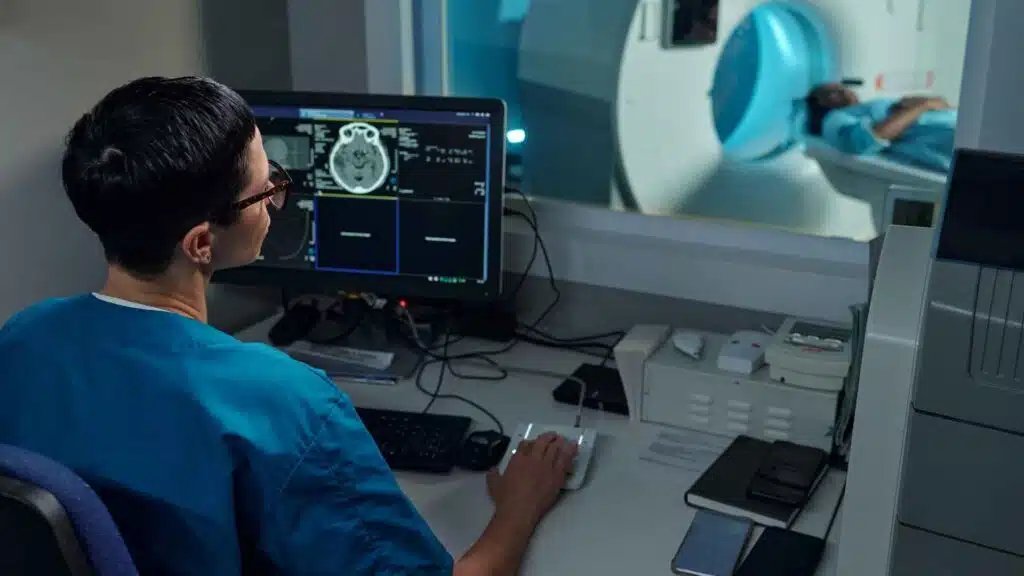
Stroke demands immediate action as every minute matters. Neuroimaging sits at the center of this race, guiding triage, ruling out hemorrhage, and unlocking thrombolysis or thrombectomy decisions. Yet misdiagnosis affects far too many stroke cases, contributing to preventable death and long-term disability at a scale that demands urgent systemic attention.

Legally Reviewed By
Attorney Rich Newsome
Catastrophic Injury & Medical Malpractice Attorney
25+ years experience

Every minute of delayed treatment destroys roughly 1.9 million neurons, according to Stroke. Accurate stroke diagnosis depends on selecting the right imaging tool at the right moment. Each modality offers a distinct trade-off between speed, availability, and diagnostic sensitivity, and understanding these differences is essential to recognizing where errors are most likely to occur.
Imaging errors in stroke fall into four overlapping categories, each capable of derailing the diagnostic process at a different point from image acquisition through final interpretation.
False negatives represent the most consequential imaging failure: a stroke is present, but the scan appears normal or inconclusive, and treatment is delayed or never initiated.
Non-contrast CT, despite its speed and availability, is fundamentally limited in the early part of an ischemic stroke. A normal CT alone cannot and should not rule out stroke for several reasons.
The base of the skull on CT scans has a blind spot. The brainstem and cerebellum sit deep inside the head, surrounded by thick bone. When a CT scanner sends X-ray beams through that much bone, it creates visual “noise” in the image. This interference can hide a stroke, even a fairly large one.
Some strokes are simply too small for a CT scan to see. Lacunar strokes happen in tiny, deep blood vessels supplying structures like the internal capsule, which controls movement, and the thalamus, which processes sensation and consciousness. These infarcts (areas of brain tissue that are dead or dying) typically fall below the detail level that a CT scanner can reliably see.
Even an MRI is not infallible. DWI-negative strokes occur in a meaningful minority of cases, particularly within the first few hours of symptom onset or when lesions are extremely small. Brainstem strokes show up disproportionately in this “missed” category because the thick bone at the base of the skull distorts MRI images there, too, not just CT scans.
Several unrelated conditions can produce brain scan appearances that look like a stroke.
Brain tumors are common offenders.
After a seizure, some patients experience temporary weakness or paralysis on one side of the body, a phenomenon known as Todd’s paralysis. This can trigger scan changes that look like a stroke, and because no one may have witnessed the seizure itself, the underlying cause is easy to miss. Severe migraines with one-sided weakness can produce scan abnormalities resembling stroke, particularly in younger patients.
Finally, certain whole-body chemical imbalances, such as dangerously low blood sugar, can alter brain appearance on MRI in ways that mimic stroke.
Interpretive errors arise not from the scan itself but from the human reading it, and they are more common than most clinicians understand.
Even a correctly ordered study can fail if acquisition is flawed.
When a stroke is missed or diagnosed too late, families are often left with difficult questions and difficult complications. If an imaging error may have played a role, it is natural to wonder whether the outcome could have been different and whether something was overlooked during the diagnostic process.
Imaging studies are important tools in identifying strokes, but interpreting those images can be complex. In some situations, signs of a stroke may be:
However, an unexpected outcome does not automatically mean negligence occurred. Determining what happened requires a careful review of the medical care provided.
Our stroke misdiagnosis lawyers can help by conducting a thorough evaluation of the circumstances surrounding your loved one’s diagnosis and treatment. This typically begins with gathering and reviewing:
We work with qualified medical experts who can assess whether the imaging was interpreted appropriately and whether the care met accepted medical standards.
The goal of this review is to provide clarity. In some cases, families find reassurance that appropriate care was provided despite a tragic outcome. In others, the evidence may indicate that preventable errors contributed to a delayed diagnosis and resulting harm.
If negligence is identified, we can explain the available legal options and help you understand the next steps. Pursuing a stroke misdiagnosis claim is always your decision. Whether you are seeking answers, accountability, or compensation for the challenges your family now faces, we are here to provide guidance, information, and support if and when you are ready.
If your loved one suffered harm after a stroke was missed or delayed, you do not have to sort through the questions alone. Our team can review the medical care that was provided, help you understand what happened, and explain any options that may be available. Whether you are seeking answers, closure, or considering legal action, we are here to provide guidance and support if and when you are ready.
Call Newsome Law whenever you are ready.
There is a question that many families of locked-in syndrome patients ask themselves, usually late at night, long after the...
Common Factors That Contribute to Misdiagnosis Stroke misdiagnosis is rarely the result of a single error. More often, it reflects...
Patients’ Rights After a Failure to Diagnose a Stroke After a stroke is misdiagnosed, patients and their families are often...
A tracheostomy and a ventilator can feel like foreign objects at first: tubes, machines, and sounds that take over a...
Locked-in syndrome (LIS) changes everything overnight, including a family's finances. Once income stops, Social Security disability benefits can become a...

Learning your child has locked-in syndrome can feel like standing in a room with no map. It's a rare diagnosis,...
While there are occasional cases that make the news of someone recovering from locked-in syndrome or a similar diagnosis, this...

The area of the brain that can cause locked-in syndrome (the pons) following a stroke is the same area of...